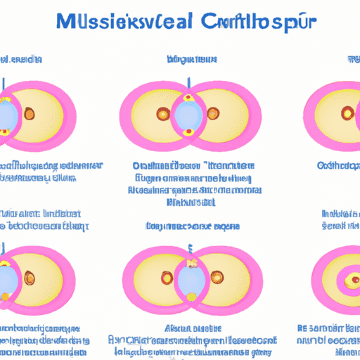
Definition of Mitosis – Does anyone feel that her face looks like her mother? Or look like your father? Or even not similar to both but more like grandfather or grandmother? How can there be similarities? Yes, there are still family blood ties. Yes, but what is the story of the similarity process?
Every child or individual must have characteristics or traits that are similar, even the same as their parents. This is because of the inheritance of characteristics or traits from parents to their children.
The inheritance of traits from parents to their descendants is called heredity.
Have you ever imagined how many cells there are in our body? Millions? or even trillions? How is it possible that we have trillions of cells when we used to come from only two cells, namely the mother’s ovum cell and the father’s sperm cell?
Turns out, cells have the ability to clone! Yes, the cells in our body are capable of doubling themselves so that we can develop, grow, even heal wounds on ourselves!
So if you have a broken heart, just calm down, your body will heal the wound by itself
It is DNA that is the gateway to heredity. The daughter cells resulting from mitosis and meiosis will have DNA in which there is genetic material from the parent cell, it may be exactly the same or a combination of both characteristics of the parent cell.
If a dividing cell is stained and examined under a microscope, dark colored chromosomes will be seen in the nucleus. Each consists of a central thread, the chromonema, which contains small swelling chromomeres that are dark in color like beads.
Some argue that the chromomeres contain genes, because early experiments clearly proved that the unit of heredity is located in the chromosomes in a linear arrangement. But the relationship between chromomeres and genes is irregular, some chromomeres have several genes and some genes are found between chromomeres.
Each chromosome, at a certain place along its body, has a clear round circular zone called a kinetochore that regulates the movement of chromosomes during cell division. Because before cell division occurs, the chromosomes thicken and become shorter, so the kinetochores become clearer and look like constrictions.
The process of inheritance of traits cannot be separated from the role of two types of cell division events, namely mitosis and meiosis.
A. Definition of Mitosis
Mitotic Division is a cell division event that produces two daughter cells with the same number of chromosomes as the parent cell. The term mitosis in a narrow sense concerns the division of the nucleus into 2 daughter nuclei and the term cytokinesis is used for the division of the cytoplasm that produces daughter cells that each contain a daughter nucleus.
You know, all cell activity is organized from the nucleus, more specifically by DNA. Therefore, the event of cell division also occurs in the nucleus. The process of cell division does not mean that it duplicates all parts of itself. However, what actually happens is that only DNA is duplicated.
Nuclear fission and cytoplasmic fission although almost always well integrated, are 2 separate and distinct processes. Mitotic division only occurs in eukaryotic cells, while prokaryotic cells cannot do it.
Why? The reason is that prokaryotic cells do not have a nucleus (cell core), cell core membrane, and mitochondria. Whereas in mitosis the organelles are involved.
In other words, the maker’s kitchen is not complete.
Where does mitosis occur? In animals and humans, the process of mitosis occurs in all body cells (somatic), except for sex cells (gametes). In animals, an aster is formed and a groove is formed at the equator on the cell membrane at the time of telophase until the two daughter cells become separated.
While in plants, mitosis occurs in the meristem network, such as the tip of the root and the tip of the stem bud. Mitotic division functions for the growth of body cells, replacing damaged body cells (regeneration), and maintaining the number of chromosomes. Mitotic division consists of nuclear division and cytoplasmic division.
Mitotic division begins with the division of the nucleus. Therefore, when we see a group of cells that are dividing, we may find one or several cells that have two nuclei. This means that the cell has finished dividing the nucleus but not yet dividing the cytoplasm.
In studying this cell division, we must know that each cell in the body has a different function. In the Cell Membrane Structure and Function Biochemistry Textbook by Andrew Johan you will better understand the function of each cell in the body.
B. Mitotic process
This process of mitosis produces two identical daughter cells, which have almost the same distribution of organelles and cell components. Mitosis and cytokinesis is the mitosis phase (M phase) in the cell cycle, where the initial cell is divided into two daughter cells that have the same genetics as the initial cell.
The main result of mitosis is the division of the genome of the initial cell into two daughter cells. The genome is made up of a number of chromosomes, which are closely coiled DNA complexes that contain vital genetic information for the proper functioning of the cell.
Because each daughter cell must be genetically identical to the original cell, the original cell must duplicate each chromosome before mitosis. So he formed himself into 2 first.
The process of DNA duplication occurs in the middle of interphase, which is the phase before the mitosis phase of the cell cycle. After duplication, each chromosome has an identical copy called a sister chromatid, which is attached to a region of the chromosome called the centromere. Sister chromatids themselves are not considered chromosomes.
C. The difference between Mitosis and Meiosis
Basically, cell division that occurs in every living being has two types, namely mitosis and meiosis. If viewed in general, the difference between mitosis and meiosis can be seen in the daughter cells produced. Mitotic cell division will form daughter cells that can divide again. Meanwhile, meiosis will form daughter cells that cannot divide again, even until fertilization. Some of the differences between mitosis and meiosis are as follows:
1. Mitotic offspring cells will be the same as the parent cells, while meiosis offspring cells will produce offspring that are different from the parent.
2. Mitosis can occur in every existing organism, but it is different with meiosis which can only occur in animals, fungi, humans, and animals.
3. The number of chromosomes of mitosis offspring is the same as its parent, while the number of chromosomes of meiosis offspring and its parent is half different.
In order to better understand the process of DNA duplication, DNA cloning, and various basic analyzes of DNA technology in biotechnology that are currently popular, Reader can read the book Fundamentals of Genetic Science by Zairin Thomy.
D. Examples of Mitosis
A type of cell division capable of producing 2 genetically identical daughter cells. That is, the two offspring cells formed have the same genetic makeup as the parent.
Almost all living things undergo the same process of mitosis, except for prokaryotes (living things that do not have a true nucleus) such as bacteria, viruses and blue algae.
Mitotic division occurs during embryo development and growth, in the replacement of worn-out cells such as blood cells, skin, intestinal lining and so on, and in wound healing.
E. Purpose of Mitotic Division
Mitotic division has three purposes, namely growth, repair, and reproduction. Explanation of the purpose of mitosis as follows.
1. Growth
All living things will definitely experience growth caused by the large number of cells that come together. Therefore, when the cells continue to increase, it will affect the living being itself.
2. Improvements
Not only is it meant to grow, but mitosis is meant for repair. In this purpose, repair will occur when the network in the living being is experiencing damage to the network.
3. Reproduction
Every living thing will definitely multiply or it can be said that it will reproduce. The reproduction process that occurs in living things definitely requires sex cells that meet each other. From that cell meeting, cell division will occur.
F. Characteristics of Mitotic Division
2. The number of daughter cells produced from mitosis is two
3. The number of chromosomes in a child is the same as the number of chromosomes in the parents, namely 2n (diploid)
4. The properties of daughter cells are the same as parent cells
5. Occurs in body cells (somatic cells), for example in embryonic tissue, including root tip, stem tip, cambium circle.
6. The purpose of mitosis is to multiply cells such as growth or to repair damaged cells.
7. Go through the stages of division, interphase, prophase, metaphase, anaphase, and telophase, but in general these stages will return to form the cell cycle.
G. Phases/Stages of Mitotic Division
At this stage of mitosis, it consists of four stages, namely prophase, metaphase, anaphase, and telophase. Before entering the 4 stages of mitosis, the cell will pass through the interphase stage first. The full explanation is below.
Interphase
However, every time the fourth phase of this phase begins, there is a preliminary term called the preliminary phase or interphase. This interphase is also often referred to as preparation for cleavage.
In the interphase, there is a process of preparation and accumulation of energy by the cell to do division. Well, what you need to know is that this process takes a very long time compared to other phases, lol.
At this stage, the cell core (nucleus) and the cell core child (nucleus) will be clearly visible. However, the chromosomes in the cell are not even visible, why is that? This is because the chromosomes are still in the form of chromatin. Chromatin is a fine thread composed of several molecules, such as RNA, DNA, and Protein.
Meanwhile, on the outside of the cell nucleus there are centrosomes. Centrosomes are cell organelles that have the function of maintaining the number of chromosomes, the number of chromosomes between the parent cell and daughter cells so that the number remains the same when cell division occurs.
So, if in animal cells, each centrosome will contain a pair of centrioles shaped like a small cylindrical body
The interphase stage is grouped into three, namely G1 phase (first gap), S phase (synthesis), and G2 phase (second gap).
a. G1 phase
The G1 phase is also called the phase of cell development and growth. This phase is marked by the development of cytoplasm (cell fluid), cell organelles, as well as the synthesis of materials that will be used in the next interphase level, which is the S phase.
At this stage, the cells grow larger. Some cell growth, including
Ada
- enlargement of nucleus size;
- increased cytoplasmic volume;
- DNA formation;
- formation of enzymes for DNA replication;
- the formation of proteins through the process of protein synthesis (transcription and translation) to drive nuclear division;
- spindle thread formation.
Subphase G-1 is the longest process in interphase, which is around 12 – 24 hours.
b. Phase S
In the S phase, there is duplication or replication of DNA as genetic material that will be passed down to daughter cells, so that two copies of DNA will be produced later. At the synthesis level, there is DNA replication along with histone proteins whose strands are called chromatin threads.
The replication process of this chromatin thread forms twins called chromatids. These two twin chromatids are attached to one centromere. The synthesis process at this level takes about 6 to 8 hours
c. G2 phase
The third phase, namely the G2 phase, DNA replication is complete. There is an increase in protein synthesis as the final stage of cell preparation to divide.
In the secondary growth stage, cell organelles and also RNA are formed. This stage takes about 3 to 4 hours and is the last process before the cells are really ready to divide.
Chromatin in the phases of the cell cycle:
- double stranded DNA
- Chromatin (single-stranded DNA with histone proteins)
- Chromatin at interphase (blue) and centromere (red)
- Compact chromatin during prophase
- Chromosomes in metaphase
At the end of interphase, a cell has a nucleus with two nucleoli (nuclei). As explained earlier, inside the nucleus there are chromatids, which are chromatin threads that have been duplicated.
In the book Encyclopedia of Biology Volume 5: DNA, RNA & Chromoson Genetics by James Bodick Dkk, Reader will learn and deepen everything about the DNA that exists and organizes the human body.
Meanwhile, outside the nucleus, centrosomes are also duplicated and will later help the process of cell division in the mitosis phase. Further, after all preparations are completed, the cell will enter the mitosis phase which consists of four stages.
1. Prophase Stage
Come on, let’s go into the initial stage of cell division, which is the prophase stage. At the beginning of prophase, the centrosomes undergo replication, resulting in two centrosomes. Then, each centrosome will move towards the opposite poles of the cell nucleus.
At almost the same time, microtubules begin to appear between the two centrosomes. These microtubules are long protein fibers that extend from the centriole in all directions.
Over time, the microtubules will form similar coils of thread that we can call spindle threads. At this stage as well, the chromatin threads begin to thicken and form chromosomes. Well, this chromosome has two identical chromatids attached to the centromere (head of the chromosome).
Well, each centromere has two kinetochores which are protein formations and eventually become the attachment point for the spindle threads. At the end of the prophase stage, the nucleus and nuclear membrane of the cell begin to disappear. In addition, the centrosomes have arrived at their respective poles.
The spindle threads will stretch from one pole to the other. This spindle thread has the role of pulling chromosomes to the middle of the cell nucleus at the next stage.
The conclusion in this phase is what happened
- Chromosomes have doubled alias become 2, then compact
- The nuclear membrane begins to break down into small parts (fragments)
2. Metaphase stage
At this stage, the nucleus and nuclear membrane of the cell become invisible. Each kinetochore at the centromere is connected to a centrosome by spindle threads. Well, later the chromatid pair moves to the center of the cell nucleus (equatorial plane) and the metaphase plate is formed.
The position of chromosomes located in the middle of the cell nucleus means that the number of chromosomes can be counted accurately and the shape of chromosomes can also be clearly observed. In this phase, Chromosomes have doubled, then compacted. The nuclear membrane begins to break down into small parts (fragments)
3. Anaphase stage
At anaphase, chromatid separation marks this phase. Starting from the centromere which then forms a new chromosome. Each chromosome is pulled by spindle threads moving towards poles in different directions. The number of chromosomes that go to one pole will be exactly the same as the number of chromosomes that go to the other pole.
So, at the end of anaphase, the chromosomes have almost reached their respective poles. In addition, cytokinesis also begins to occur. What is cytokinesis? Cytokinesis is the phase of division or separation of cytoplasm, organelles, and cellular membranes. This division starts from the edge of the cell (cell membrane) moving towards the center of the cell, so that it will produce two cells called daughter cells.
So in this phase it is concluded that:
- Chromosomes move towards opposite poles.
- At the end of anaphase both poles of the cell have the same number of chromosomes
4. Telophase stage
Well, the next stage is telophase. At this stage we have entered the final stage of mitosis. At this stage, the chromosomes have arrived at their respective poles.
The spindle threads begin to disappear and the cell’s nuclear membrane also begins to exist between the two separate sets of chromosomes.
In this phase:
Chromosomes gradually thin and change shape into chromatin threads again.
- The nuclear membrane begins to rejoin
- Two daughter cells are formed which are diploid
The process of mitosis produces 2 daughter cells from 1 original cell. Because all cells in the body originate from the mitosis of a fertilized egg cell, each cell has the same type and number of chromosomes, and the same number and type of genes.
The speed and frequency of cell division varies greatly in different tissues and in different animal species.
At the stage of embryonic development the interval between cell divisions may only be around 30 minutes. In certain adult tissues, especially nerve cells, cell division is very rare.
In other adult tissues such as the spinal cord which is where red blood cells are produced, cell division must occur frequently to provide the 10,000,000 red blood cells produced every second of the day and night by humans.

